
Figure 8.
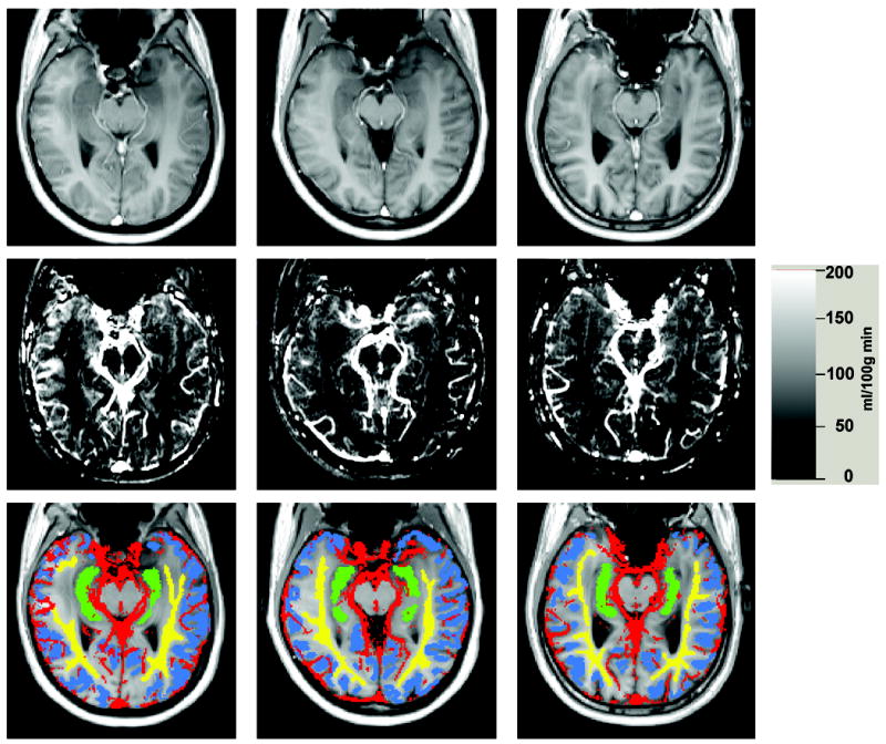
Left column: 30 year old woman, middle column: 51 year old woman, right column: 68 year old man. Top row: tagged TrueFISP images. Middle row: CBF images. Bottom row: tissue ROIs used in this study: yellow=white matter ROI, blue = cortical gray matter ROI, green=hippocampal ROI, red=vascular ROI.