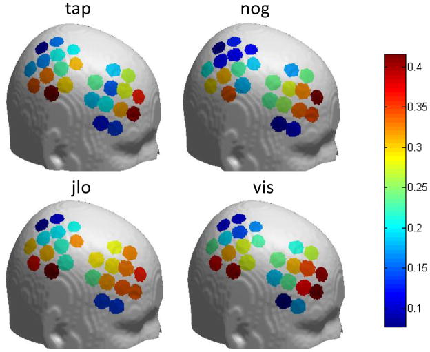
Figure 6.
fMRI-oxy-Hb correlation for each channel in different tasks. In all four tasks, the correlation is higher in the middle frontal area and the inferior parietal area than other areas. The location of each channel for individual participants was mapped to a standard brain, and then a single position for each channel was calculated by averaging across the 13 participants. tap - Tapping of fingers on left hand; nog - Go/no-go; jlo - Judgment of line orientation; vis - Visuospatial N-Back working memory task.

