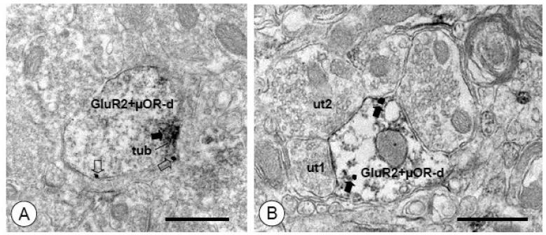
Figure 8. Small dendritic processes of CeA neurons express both GluR2 and μOR.
(A). A small dendritic profile (GluR2+μOR-d) shows immunoperoxidase labeling for GluR2 (arrow) and immunogold labeling for μOR (open arrows). A dense aggregation of immunoperoxidase reaction product for GluR2 is seen near the plasma membrane and an associated tubulovesicular organelle (tub). Immunogold particles for μOR are also present on the extrasynaptic plasmalemma. (B). A small dual labeled dendritic profile (GluR2+μOR-d) is contacted by unlabeled axon terminals (ut1, ut2). A postsynaptic specialization, which also appears to express immunoperoxidase reaction product (arrows), is formed by one of these terminals (ut1). Immunogold μOR labeling (open arrows) is present on the nearby extrasynaptic membrane as well as intracellularly. Scale Bars= 0.5 μm

