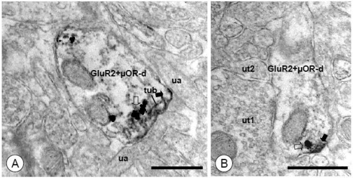
Figure 9. GluR2 and μOR labeling are found in closely apposed subcellular structures in dendritic profiles of CeA neurons.
(A). A dual labeled dendritic profile (GluR2+μOR-d) is contacted by unlabeled axons (ua). Immunoperoxidase labeling for GluR2 (filled arrow) is present near the surface membrane, as well as an adjacent tubulovesicular organelle (tub). A cluster of immunogold particles for μOR (open arrow) is present on an adjacent membrane. (B). A dendritic profile shows discrete immunoperoxidase reaction product for GluR2 (arrow) near the plasma membrane. A single immunogold-silver particle for μOR (open arrow) is present in an adjoining membrane. Unlabeled axon terminals (ut1, ut2) are adjacent to this dendrite. Scale Bars= 0.5 μm

