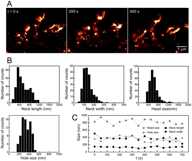
Figure 4. Spine dynamics in mature hippocampal neurons.
(A) Super-resolution time-lapse imaging of dendritic spines from a neuron expressing ABP-tdEosFP at DIV 27. Each PALM image was reconstructed from 2000 frames recorded at 25 ms. (B) Quantification of morphological spine parameters (as in Fig. 2E) in living neurons (neck width: 140±45 nm mean ± standard deviation, N = 236 spines; neck length: 550±240 nm, N = 385; head diameter: 600±180 nm, N = 385; diameter of holes and cup-shapes: 340±85 nm, N = 81; from 12 cells and 3 independent experiments). (C) Baseline dynamics of the morphological parameters of an individual spine under control conditions.

