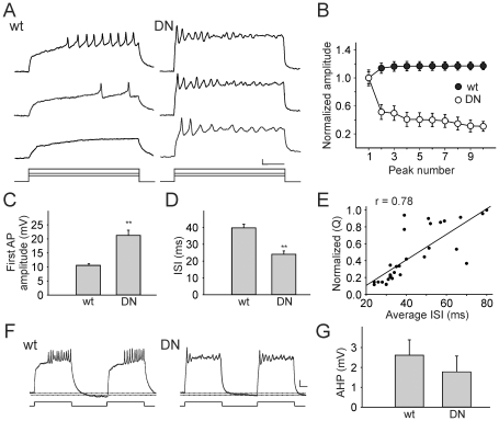
Figure 4. Kv4 is Required for Maintaining Excitability in Drosophila Neurons.
A, Representative firing patterns recorded with current injections of 40, 60, and 100 pA from wild-type (wt) and DNKv4 (DN) neurons. With increasing current injections, wt neurons display a reduction in the delay to the first AP and an augmentation in AP firing frequency, maintaining excitability for the duration of the 500 ms stimulus. DN neurons display little delay to the first AP, and subsequent peaks decrease in amplitude, compared with wt. B, Plotted are amplitudes of the first 10 peaks, representing APs or graded potentials, normalized to the first AP (N = 30 for wt, N = 20 for DN); a pronounced adaptation of peaks in DN neurons is observed. C, The average amplitude of the first AP in DN neurons (N = 20) is significantly greater than wt (N = 30). D, The interspike interval (ISI), measured as the time between the peaks of the first and second APs, is significantly decreased in DN neurons (N = 20) compared to wt (N = 30). E, Current-clamp recording was performed (100 pA, 500 ms), followed by a voltage-clamp recording to isolate the Kv4 current, as described in Figure 1A. Shown is the normalized charge carried by the Kv4 current plotted against the average interspike interval between each AP fired during the stimulus. A positive correlation (r = 0.78, N = 15) is seen between Kv4 current charge and average ISI. F–G, Shown are voltage responses of representative wt and DN neurons to a pair of 500 ms current injections of 100 pA, with an interval of 500 ms. The AHP that follows each stimulus “burst” is measured as the hyperpolarization beyond the membrane potential before the stimulus, as indicated by the doted lines. No significant difference in this “interburst” AHP was seen between wild-type and DNKv4 neurons (N = 9 for wt, N = 11 for DN) (G). Scale bars represent 10 mV and 100 ms.

