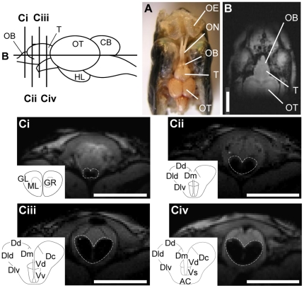
Figure 4. Magnetic resonance imaging (MRI) scan and dissected view of the front part of a lacustrine sockeye salmon head.
A: Dissected view; B: MRI horizontal section; Ci–iv: serial MRI scan of frontal sections of the olfactory bulb (Ci) and the telencephalon (Cii–iv). The olfactory bulb and telencephalon are outlined by dotted lines. Scale bars = 5.0 mm. AC, anterior commissure; CB, cerebellum; Dc, central area of dorsal telencephalon; Dd, dorsal area of dorsal telencephalon; Dld, dorsal part of lateral area of dorsal telencephalon; Dlv, ventral part of lateral area of dorsal telencephalon; Dm, medial area of dorsal telencephalon; GL, glomerular layer; GR, granular layer; HL, hypothalamus; ML, mitral cell layer; OB, olfactory bulb; OE, olfactory epithelium; ON, olfactory nerve; OT, optic tectum; T, telencephalon; Vd, dorsal area of ventral telencephalon; Vs supracommissural area of ventral telencephalon; Vv, ventral area of ventral telencephalon.

