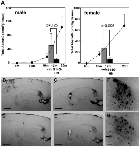
Figure 4. Brain Abeta level was sustained in S14G-HN-treated 3xTg-AD mice.
After behavioral tests, mice were sacrificed at 17 months of age. Abeta was extracted from brain homogenates using formic acid. A. Amount of human Abeta40 was measured by ELISA and shown as mean±SEM. P values of Student t-test are shown. Number of animals were vehicle male = 7, female = 9, S14G-HN male = 9, female = 7. Lower level of Abeta was detected in S14G-HN treated mice (black columns). Statistical significance was observed in female but not male compared with vehicle treated mice (dark grey columns). Dotted line plots show Abeta40 levels of naïve 3xTg-AD mice at the indicated age [37]. B–G. Immunohistochemical analysis of amyloid plaques. Brains were fixed with 4% paraformaldehyde and 30 µm sagittal sections were subjected to immunostaining using anti-Abeta antibody (82E1) and biotin-conjugated secondary antibody followed by visualization with ABC method. B, D, F. vehicle-treated female mice, C, E, G. S14G-HN-treated female mice. F, G. magnified view of subculum regions in C and E, respectively. Bar = 500 µm in B-E, = 100 µm in F, G.

