Figure 1.
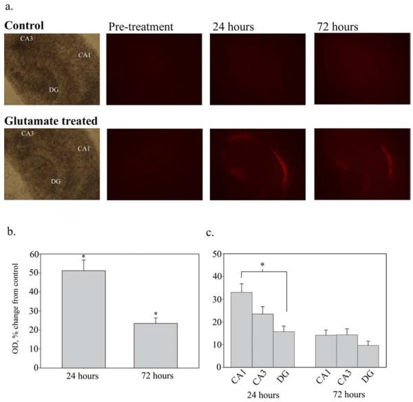
A. Propidium Iodide uptake - Propidium Iodide (PI) uptake in OHSCs following 35 minute treatment with 3.5mM glutamate. PI staining indicates increased cell death in glutamate treated slices as compared to controls at both 24 and 72 hours following treatment. Note that significant cell death is observed in the CA1 cell region. (4X magnification). B. Comparison of PI uptake - Quantification of PI uptake in OHSCs at 24 and 72 hours after injury, expressed as a percent change from control. PI uptake was measured as mean optical density of the whole slice and normalized to age-matched controls. PI staining indicated a significant increase in cell death at both 24 (n=121, 174 for control and glutamate treated, respectively) and 72 hours (n=108, 154 for control and glutamate treated, respectively) after glutamate treatment (*p<0.001, Mann-Whitney Rank Sum test). C. PI uptake by cell region - At 24 hours, cell death is significantly higher in the CA1 cell region than it is in the dentate gyrus (DG) (*p<0.05, Kruskall-Wallis One Way ANOVA On Ranks). The CA3 region did not have a significant difference in PI uptake from either the CA1 or DG. At 72 hours, there is no significant difference in PI uptake between cell regions. (n=154)

