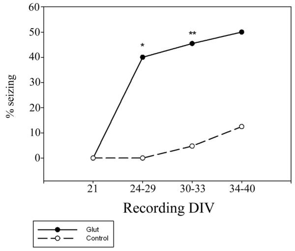
Figure 5.
Development of seizures after injury - After glutamate injury (solid line), a significant percentage of OHSCs display seizure activity in field potential recordings as compared to age matched controls (dashed line). This change is long lasting and was observed up to 40 days in vitro. Within hours of injury (DIV 21), OHSCs did not display seizure events in either the control or injured groups (n=6 each). Between 3 and 8 days after injury (DIV 24-29), 40% of injured OHSCs and 0% of controls displayed seizure events (n=10 each, *p<0.05, Fisher’s exact test). In our established experimental time frame, DIV 30-33, we observed seizure events in 46.25% of glutamate injured OHSCs and 7.14% of controls (n=80, 28, **p<0.001, Chi-square analysis). By DIV 34-40, seizure events were observed in 50% of injured slices and 12.5% of controls (n=8, 9).

