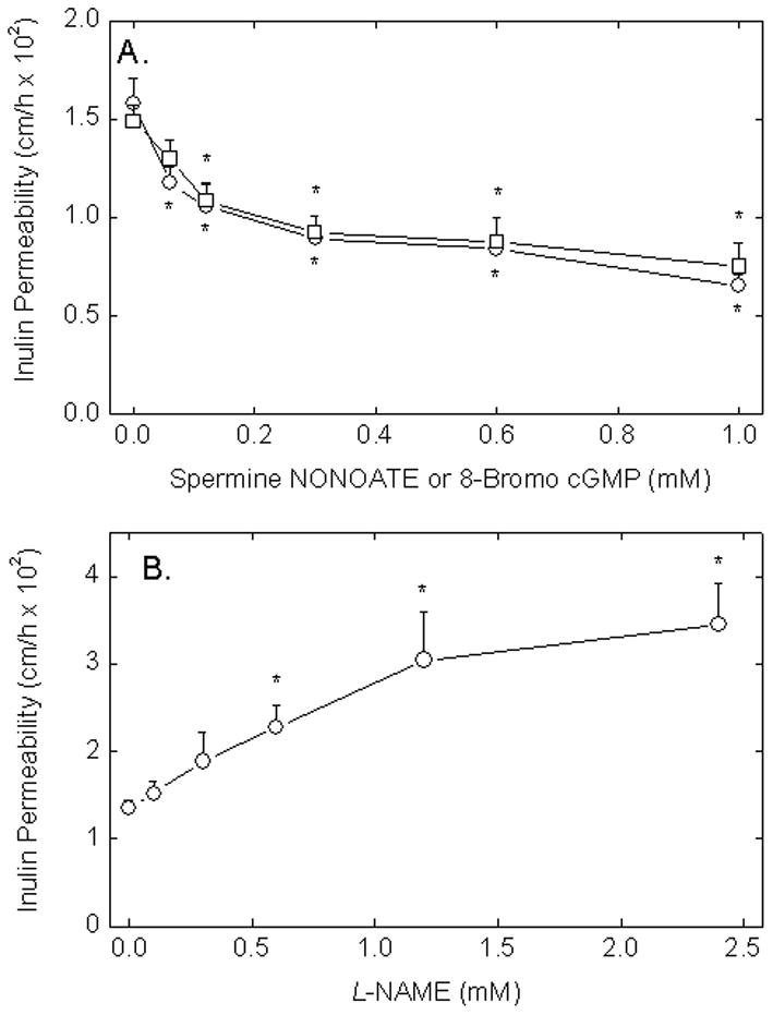
Figure 1. eNOS-derived NO decreases endothelial barrier permeability in EA.hy926 cells.

Panel A. Cells in culture on porous membrane filters were incubated at 37 °C with increasing concentrations of spermine NONOATE (circles) or 8-bromo-cGMP (squares). Spermine NONOATE was added just before the 60 min inulin transfer assay due to its instability, whereas cGMP was added 30 min before the assay to allow it to enter cells. The transfer assay was carried out as described in Materials and methods. Panel B. Cells were treated at 37 °C with increasing concentrations of L-NAME for 30 min followed by the 60 min inulin transfer assay. Results for each panel are shown from 3–5 experiments, with an “*” indicating p < 0.05 compared to the untreated sample.
