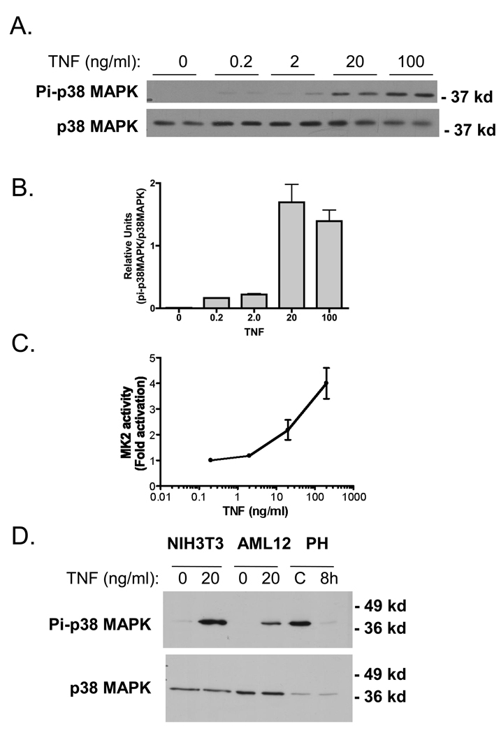
Fig 6.
Stimulation of p38 MAPK and MK2 by TNF in vitro. A) Western blot analysis of p38 MAPK activation with increasing concentrations of TNF for 10 min. in AML12 cells. B) Densitometry analysis of activated p38 MAPK with increasing concentrations of TNF. C) MK2 activation with increasing concentrations of TNF using the MK2 immunoprecipitation kinase assay described in Materials and Methods. MK2 activity is presented as fold activation (to represent the results of 3 independent experiments.). D) Western blot analysis of p38 MAPK activation in AML12 and NIH3T3 treated with 20 ng/ml for 10 min. Protein lysates from whole mouse liver from non-surgical (−) and 8hr post 2/3 PH (8h) are included for comparison.

