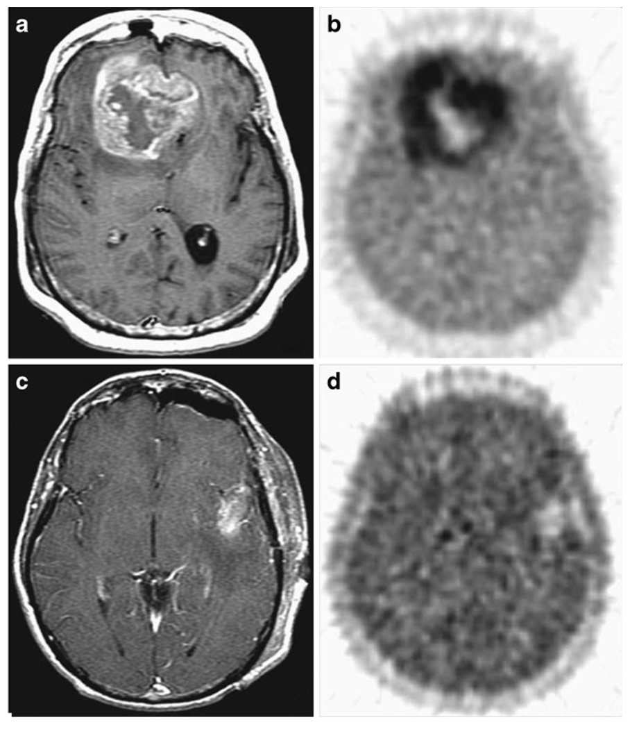
Fig. 6.
a, b A patient with bifrontal glioblastoma multi-forme, with MRI demonstrating a 20 cm3 mass with a necrotic core, and 18F-FMISO image in the same plane with a T/Bmax ratio of 3.0. c, dA separate patient with left temporal glioblastoma multiforme tumor 7 cm3 in volume after total gross resection. 18F-FMISO had a T/Bmax ratio of 1.7. Reprinted with permission from the American Association of Cancer Research [162]

