Fig. 3.

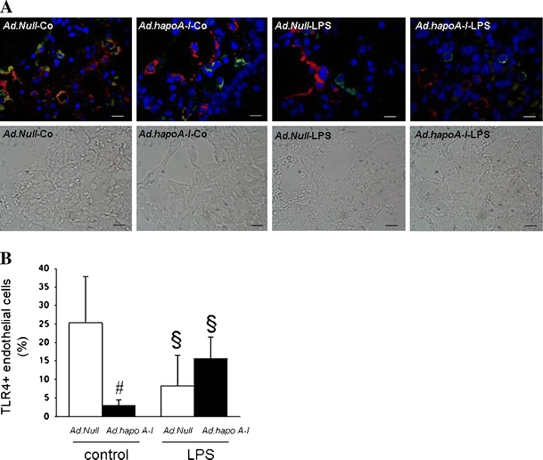
Apo A–I transfer reduces endothelial TLR4 expression. Endothelial TLR4 expression in the lung of C57BL/6 mice 14 days after intravenous injection with Ad.Null or Ad.hapoA–I and 20 h after intraperitoneal injection with saline or LPS. a Representative fluorescent microscopy pictures of double-stained TLR4 (green)/von Willebrand factor (red) cryosections of (from left to right) Ad.Null-control, Ad.hapoA-I-control, Ad.Null-LPS and Ad.hapoA-I-LPS mice with below respective phase contrast pictures (magnification ×1,000; bar corresponds to 10 μm). b Bar graph representing per cent of endothelial cells expressing TLR4 represented as mean ± SEM (n = 5–7), with Ad.Null (open bars) and Ad.hapoA-I (black bars). # p < 0.05 versus Ad.Null-control, *p < 0.05 versus Ad.Null-LPS, § p < 0.05 versus controls
