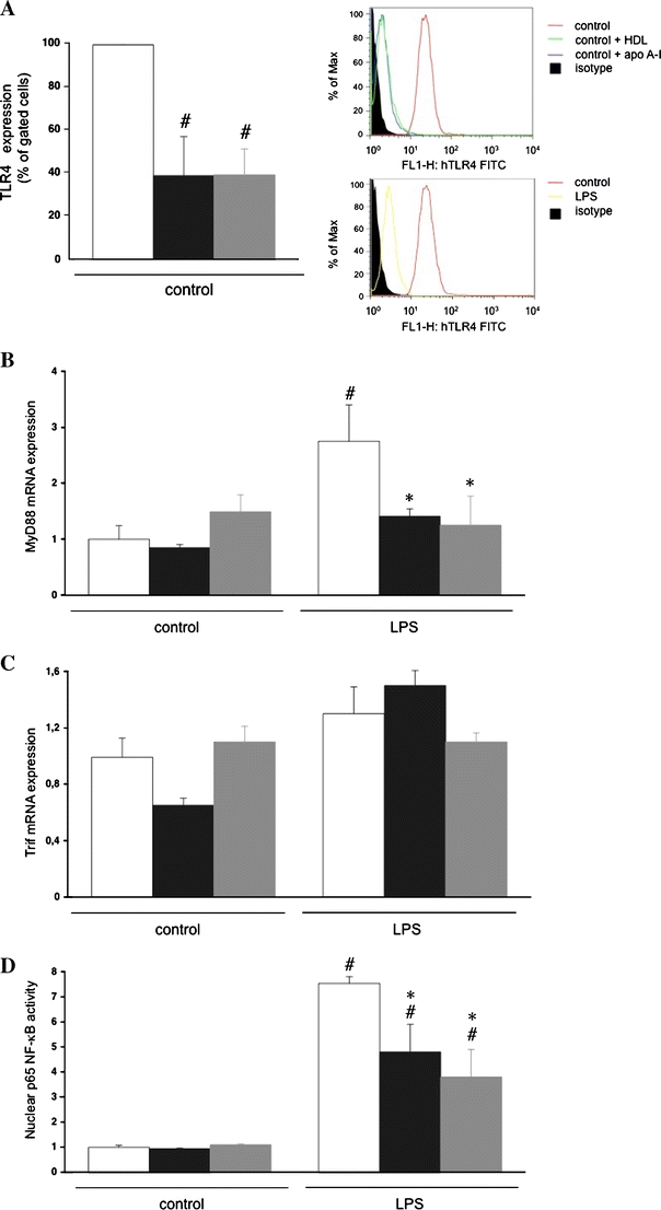
Fig. 4.

HDL and its component apo A–I reduce TLR4 expression and underlying signalling in human microvascular endothelial cells-1. Human microvascular endothelial cells-1 (HMEC-1) were incubated in the presence or absence of HDL (50 μg/ml) or apo A–I (35 μg/ml) for 24 h. Next, LPS (100 ng/ml) was supplemented in the absence of HDL or apo A–I for 2 h for FACS and mRNA expression or for 4 h for NF-κB activity analysis, respectively. a Bar graph representing per cent gated cells of n = 3 independent experiments. Right upper panel Histogram representing counts of human TLR4 expression in control, control+apo A–I, control+HDL and isotype control (see legend on the right). Right lower panel Histogram representing counts of human TLR4 expression in control, LPS and isotype control (see legend on the right). Bar graphs representing MyD88 mRNA expression (b), TRIF mRNA expression (c) and p65 NF-κB activity (d), relative to the normal control group set as 1. Bar graphs indicate mean ± SEM (n = 4), with untreated (open bars), HDL (black bars) and apo A-I (grey bars). # p < 0.01 versus untreated control, *p < 0.05 versus untreated LPS
