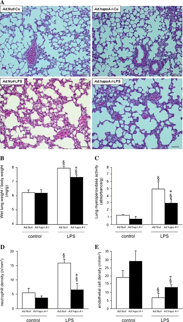
Fig. 5.

Apo A–I transfer attenuates LPS-induced lung damage. a Representative photomicrographs of haematoxylin and eosin-stained paraffin sections (magnification ×200; bar corresponds to 100 μm) from lungs of control C57BL/6 mice 14 days after intravenous injection with Ad.Null (Ad.Null-Co) or Ad.hapoA-I (Ad.hapoA-I-Co, top) and C57BL/6 mice 20 h after LPS treatment (Ad.Null-LPS and Ad.hapoA-I-LPS, bottom). Extensive interstitial oedema, infiltration of inflammatory cells and haemorrhage is present in Ad.Null-LPS mice, whereas lung damage is markedly reduced in mice overexpressing human apo A–I compared with Ad.Null-LPS mice. Bar graphs representing wet lung weight-to-body weight ratio (b), lung myeloperoxidase activity (c), as marker of neutrophil infiltration, neutrophil cell density (n/mm2) (d) and endothelial cell density (n/mm2) (e). Bar graphs indicate mean ± SEM (n = 6), with Ad.Null (open bars) and Ad.hapoA-I (black bars). *p < 0.05 versus Ad.Null-LPS, § p < 0.05 versus controls
