
Fig. 1.
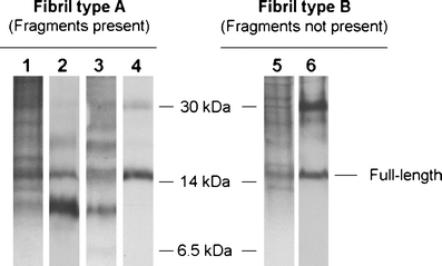
The two fibril composition types, shown by SDS-polyacrylamide gel electrophoresis (PAGE) and Western blot analyses on adipose tissue. Lanes 1–4 show a fibril composition of both full-length and fragmented ATTR (fibril type A) while lanes 5–6 show a fibril composition of only full-length ATTR (fibril type B). Lanes 1 and 5 = Coomassie Blue-stained SDS-PAGE gels; lanes 2 and 6 = Western blot using an in-house produced antiserum against TTR50–127 that detects C-terminal fragments; lane 3 = Western blot using an in-house produced antiserum against TTR24–35 that detects fragments containing position 30; lane 4 = Western blot using commercial α-TTR antibodies (Dako), which do not recognize the fragmented ATTR species [12]