
Fig. 1.
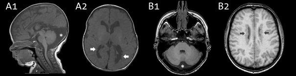
Neuroimaging characteristics of a child (patient 2) in A1 and A2, and an adult (patient 5) in B1 and B2. All are T1 weighted MRI images. Note the periventricular nodular heterotopia (denoted by arrows) and the enlarged retrocerebellar space (denoted by a star)