Figure 5.
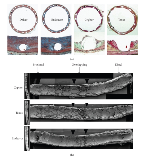
(a) X-rays of longitudinally cut rabbit iliac arteries at 21 days after placement of overlapping ZES, SES, and PES. The extent of stent coverage by endothelial cells was greatest with ZES, with almost complete coverage in the proximal and distal segments and significantly greater coverage in the overlapped segment compared with SES and PES. (b) Photomicrographs showing the amount of neointimal thickness at 28 days after placement of Endeavor zotarolimus-eluting stents (ZESs), Cypher sirolimus-eluting stents (SESs), Taxus paclitaxel-eluting stents (PESs), and Driver bare metal stents (BMSs) in rabbit iliac arteries. With SES, there were focally uncovered stent struts, which were associated with inflammation consisting of heterophils or eosinophils and giant cells. Adapted from [49].

