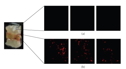
Figure 6.
Transplanted BMC migrated into mucosal damaged area. 1 × 10⁷ BMC were labeled using Vivotag750. These cells were transplanted into AA + 5-FU model via tail vein. After 24 hours, mice were sacrificed, and the colon was observed. The labeled cells could be detected in mucosal damaged area by fluorescence microscope. Transplanted BMC accumulated significantly in mucosal damaged area (b), but not in normal area (a). Three pictures were z-axis high, middle, and low in one of the particular area.

