
Figure 2.
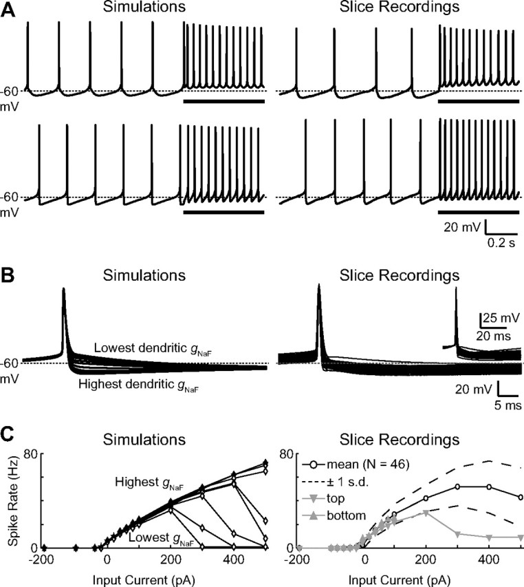
Realistic GP neuron physiology is observed for a wide range of dendritic gNaF levels. A, Simulation traces of spontaneous and current-driven (+100 pA into the soma during the underlined periods) spiking are shown for the model neurons with gNaF gradient constants of 75 μm (top left) and 250 μm (bottom left). The most apparent difference between the two was in the shape of the AHP following each spike. Voltage traces from two GP neuron brain slice recordings are shown (right) simply to illustrate the point that real GP neurons can have more rounded or sharp AHP shapes resembling the different model neurons. B, A comparison of spontaneous spike shapes between all of the model neurons revealed a direct connection between the dendritic gNaF level and the depth of the spike AHP, with a higher dendritic gNaF level resulting in a deeper spike AHP (left). Slice recordings of spontaneous action potentials from 46 GP neurons show that the set of model neurons spanned the whole range of typical GP neuron variability in this characteristic (right; to show AHP variability more directly, the inset shows the same traces vertically shifted so that all were at −50 mV 2 ms before the peak of the spike). C, The frequency responses of the nine model neurons to somatic current injections were very similar for current amplitudes between −200 and +200 pA (left). For larger positive current injections, model neurons with lower dendritic gNaF were more prone to depolarization block. GP neurons from slice recordings showed a similar average frequency-current response and also varied in their tendency to enter depolarization block at higher current levels (right). Some recordings do not show data points above +100 pA current injection because such data were not obtained for these neurons.