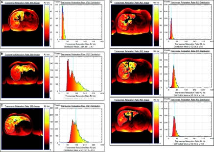
Figure 1.
Liver R2 images and distributions for four subjects with different degrees of iron (Fe) overload and pathologic conditions: (A) healthy control, (B) hereditary hemochromatosis, (C) ESKD patient with >6 g cumulative Fe dose, and (D) Fe deficient CKD patient before and 2 and 12 weeks after 1 g intravenous Fe (top to bottom). Note that the liver R2 images are superimposed on standard spin-echo images for registration purposes. Note that to enable visualization of the heterogeneity of R2 within each liver, the color scale within each liver is adjusted for each image such that zero corresponds to voxel R2 of zero, whereas the maximum of the color scale is scaled to the maximum R2 value within the liver.

