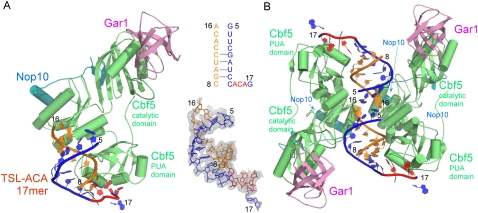
FIGURE 3.
Structure of the Cbf5-Nop10-Gar1 complex bound to TSL-ACA 17mer RNA. (A) The overall view of the Cbf5-Nop10-Gar1 complex. The 3Fo-2Fc map computed prior to modeling of the RNA is shown around the final RNA model. The ACA-binding strand is colored in blue (ACA is in red) and contains nucleotides 5–17. The complementary strand is colored in orange and contains nucleotides 8–16. (B) The packing of two symmetry related protein-RNA complexes indicates some weak interactions between the ends of the two RNA duplexes.

