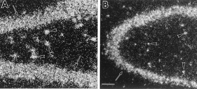
Figure 2.
Representative dark-field photomicrographs of emulsion autoradiographic analysis for GLUTx1 mRNA expression in the rat hippocampus. (A) Reduced silver grains for GLUTx1 were detected over granule neurons of the dentate gyrus (arrows) and nonprincipal cells in the hilus (arrowheads). (B) Reduced silver grains of GLUTx1 mRNA over CA3 pyramidal neurons (arrows) and nonprincipal cells (arrowheads). (Bar = 100 μ.)

