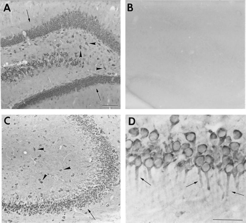
Figure 5.
Representative bright-field photomicrographs of GLUTx1 immunoreactivity in the rat hippocampus. (A) GLUTx1 immunoreactivity in granule neurons of the dentate gyrus (arrows) and nonprincipal cells of the hilus (arrowheads). (B) Preabsorption of GLUTx1 antisera eliminates GLUTx1 immunoreactivity in the hilus. (C) Low-power magnification of GLUTx1 immunoreactivity in pyramidal neurons (arrows) and nonprincipal cells (arrowheads) in CA3 of Ammon's Horn. (D) Higher-power magnification of GLUTx1 immunolabeling showing cytoplasmic labeling and GLUTx1 immunoreactivity in the proximal dendrites of CA1 pyramidal neurons (arrows). (Bars = 100 μ.)

