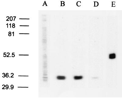
Figure 6.
Immunoblot analysis of membrane fractions isolated from rat hippocampus using GLUTx1 antisera. Lane A, GLUTx1 primary antisera (1:1,000) weakly immunodetect a 35-kDa protein in hippocampal plasma membrane fractions; lanes B and C, GLUTx1 antisera immunodetect a single protein at approximately 35 kDa in hippocampal intracellular membrane-containing fractions isolated from control rats (lane B) and STZ diabetic rats (lane C); lane D, preabsorption of GLUTx1 antisera with blocking peptide eliminates the detection of this 35-kDa protein in control cytoplasmic fractions; lane E, β-tubulin mAb (Sigma) immunodetects a single protein at approximately 52 kDa in cytoplasmic fractions, confirming the successful isolation of hippocampal cytoplasmic fractions from hippocampal plasma membrane fractions. (All lanes equal 50 μg of protein; lanes are representative of at least four separate immunoblot experiments.)

