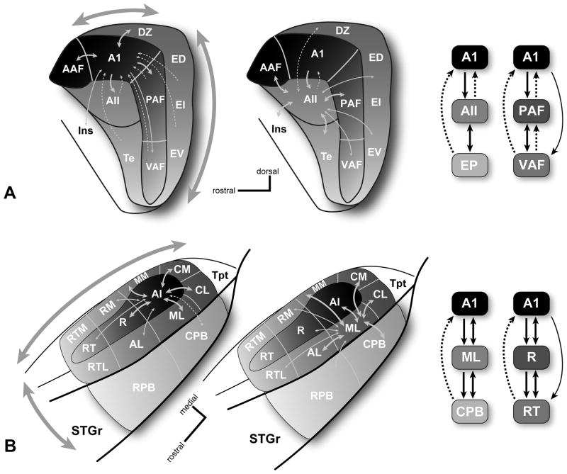
Fig. 3.
Local connections of selected core and belt areas. (A) Left, connections of the core area, A1 (left), and belt area, AII (middle), in the cat. Right, schematics of information flow along the caudal-rostral axis (A1-AII-EP)(core-belt-parabelt?) and dorsal-ventral axis in the core (A1-PAF-VAF). (B) Left, connections of the core area, A1 (left), and lateral belt area, ML (middle) in the primate. Right, schematics of information flow along the medial-lateral axis (A1-ML-CPB)(core-belt-parabelt) and caudal-rostral axis in the core (A1-R-RT). Line thickness denotes the relative density of each projection. Dashed lines indicate feedback projections. Shading intensity (all panels) and large arrows (left panels) denote anatomical and physiological gradients along two major axes of information flow in both species. See text for details.

