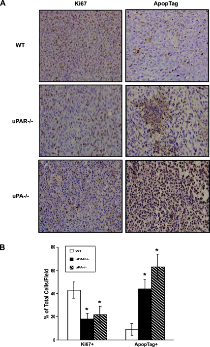
Figure 3.
Effects of uPA or uPAR deficiency on RM-1 tumor cell proliferation and apoptosis. Tumor sections were immunohistochemically stained with Ki-67 monoclonal antibody or by an ApopTag in situ detection kit, respectively. (A) Representative photographs of immunohistochemical staining. (B) Quantified data were determined by the number of Ki-67-positive cells or the number of apoptotic-positive nuclei dividing the total number of cells in five randomly selected fields under lightmicroscopy (x400). Values representmean ± SEM. *P < .001, compared with the percentage of positive cells in tumor sections from the WT mice.

