Figure 5.
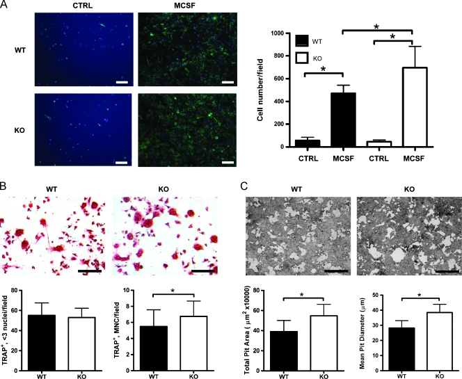
Osteoclast maturation is accelerated in SPARC KO mice. BMMΦs were isolated from the bone marrow of SPARC WT (black columns) or KO (white columns) mice. (A) Marrow was flushed and plated in the presence of 100 ng/ml M-CSF. On the third day, cells were fixed and stained for the macrophage marker F4/80 (green) to visualize BMMΦs. DAPI staining (blue) was used to show total cell numbers. Scale bars, 50 µm. F4/80-positive cells were counted and displayed as mean ± SD (n = 7 fields from triplicate experiments). *P < .05 by one-way ANOVA. (B) BMMΦ cells developed in vitro were then treated with 30 ng/ml M-CSF and 100 ng/ml RANKL for 3 more days. Scale bars, 50 µm. Cells were stained for TRAP, counted, and displayed as mean ± SD (n = 12 fields from triplicate experiments). *P < .05 by Student's t test. (C) BMMΦ cells developed in vitro were plated, in the presence of 30 ng/ml M-CSF and 100 ng/ml RANKL, on calcium phosphate-coated coverslips. Scale bars, 50 µm. Pit formation was quantified 3 days later with total pit area/field and mean pit diameter was shown as mean ± SD (n = 20 fields from quadruplicate experiments). *P < .05 by Student's t test.

