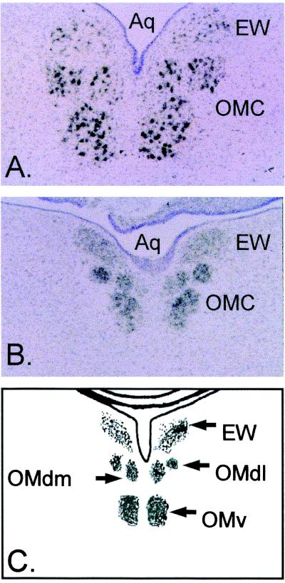
Figure 1.
Expression of β-Neu1 transcripts in developing chick oculomotor nuclei. Bright-field micrographs of in situ hybridization histochemistry with antisense riboprobes directed against EGF-domains conserved in all β-NEU1 isoforms. Sections were counterstained with cresyl violet. Note robust hybridization signal in the Edinger–Westphal nucleus (EW) and oculomotor complex (OMC) of E13 (A) and E9 (B) chick embryos. (C) The relative locations of midbrain oculomotor nuclei. The dorsal lateral oculomotor nucleus (OMdl), dorsal medial oculomotor nucleus (OMdm), ventral oculomotor nucleus (OMv), and mesencephalic aqueduct (Aq).

