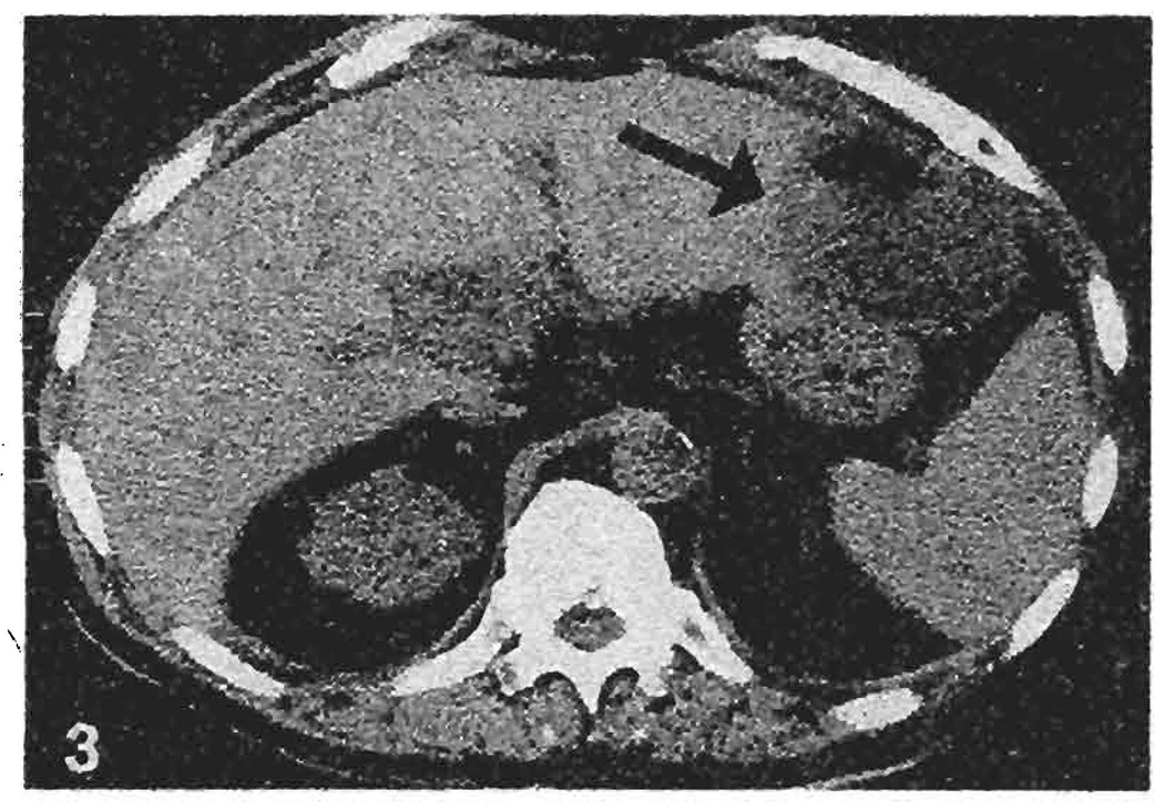
Fig. 3.
Computed tomogram of case 2 at 6 months following orthotopic liver transplantation. An intrahepatic fluid collection with an air-fluid level measuring 6.0 × 6.0 cm is visible in the left lateral segment of the transplant liver (arrow). Aspiration of the collection yielded 120 cc of pus, which grew enterococcus and diphtheloids

