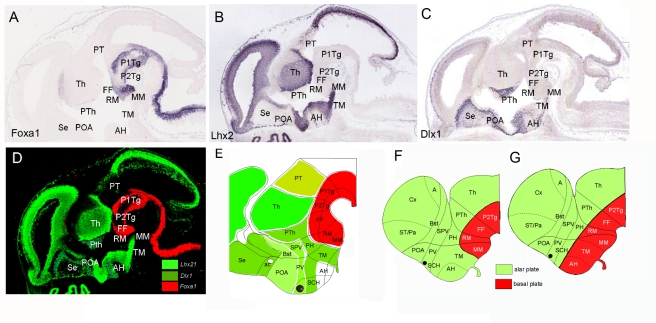
Figure 7. Combinatorial analysis of several transcription factors' patterns in the hypothalamus reveals a new model of mammalian hypothalamic organization.
(A) Foxa1 expression pattern in the basal plate of rhombencephalic, mesencephalic, diencephalic, and caudal hypothalamic neuroepithelium. This pattern is representative of other transcription factors such as Lmx1a, Lmx1b, Barhl1, Dbx1, Pax7, Olig2, Rarb, Dfp3, Lhx1, Lhx5, Irx1, and Irx3, expressed in the prosencephalic basal plate, including hypothalamus, where they were exclusively localized in the caudal regions: mammillar (MM) and/or retromammillar (RM) areas. (B and C) Lhx2 and Dlx1 expression patterns are representative of transcription factors expressed in alar prosencephalic derivatives (telencephalon, prethalamus, and thalamus) showing expression in TM and AH areas (currently described as basal plate hypothalamic domains), as well as in alar hypothalamic regions such as the suprachiasmatic (SCH), paraventricular (PV), and supraopto-paraventricular (SPV) areas. These patterns are representative of other genes expressed in alar derivatives including the TM and AH regions: Lhx6, Lhx9, Dlx1, Dlx2, Dlx5, Unc4, Cited, Rorb, Arx, Foxa2, and Otx2. (D) Photoshop composition to illustrate the alar expression patterns of Lhx2 and Dlx1 (green) and the Foxa1 basal expression (red). (E) Schematic representation of the analyzed patterns suggesting that the mammillar and retromammillar areas show basal plate molecular characteristics, while the TM and AH regions showed alar plate molecular characteristics. (F and G) Representation of the new revised topologic model that incorporates the TM and AH regions into the alar plate (F), compared to the currently accepted prosomeric model (G).The merged color composites are the product of alignment, superposition of sections, and editing using a computer program. A detailed description of the methods used to obtain such figures is included in Text S1. A, amygdale; ac, anterior commissure; Bst, bed nucleus of stria terminalis; Cx, cortex; FF, forel fields; P1Tg, pretectal tegmentum; P2Tg, thalamic tegmentum; PH, posterior hypothalamic area; POA, preoptic area; PT, pretectum; PTh, prethalamus; PV, paraventricular; SCH, suprachiasmatic; Se, septum; SPV, supraopto-paraventricular; ST/Pa, striatum/pallidum; Th, thalamus.

