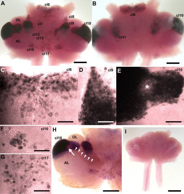
Figure 4.
In situ hybridization for 5-HT1α mRNA in whole mount brains (n = 6) of P. clarkii. (A-H) Labeling for the 5-HT1α transcript with the antisense riboprobe. (A) Ventral view of the brain after hybridization. (B) Dorsal view of the brain after hybridization. Cell clusters (cl) positive for hybridization signals were darkly stained. Selected cell clusters are more highly magnified in (C-G). Intense 5-HT1α message labeled with anti-sense riboprobe (purple to black) is found in soma clusters 6 (C), 9 (D), 10 (E), 16 (F), 17 (G), and 7, 8, 11, 12, 13 (not shown). (I) The sense probe indicated no detectable signal. (H) In some preparations, a lower 5-HT1α expression area was found (arrow) in the area where the glutamine synthetase-labeled migratory stream (brown, indicated with arrowheads just above the stream) inserts into cluster 10. Scale bars: 300 μm (A, B, H); 100 μm (C-G); 500 μm (I). AL, accessory lobe; OL, olfactory lobe. The asterisk in (E) marks the area in cluster 10 with reduced 5-HT1α mRNA expression.

