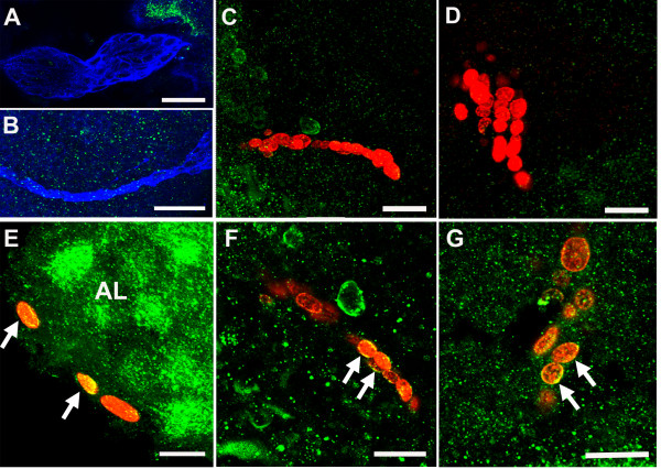
Figure 8.
Double immunofluoresence for 5-HT1α (green)/BrdU (red) (n = 6 brains), or 5-HT1α (green)/glutamine synthetase (blue) (n = 4 brains), reveals the expression of 5-HT1α protein in S phase cells in the distal streams near the proliferation zones, and in the proliferation zones themselves. (A, B) Cells in the neurogenic niche (A) and proximal and medial parts of the migratory streams (B) do not label for the receptor. (E-G) Some cells in the distal end of the stream near the proliferation zones (E), as well as cells in the proliferation zones of cluster 9 (F) and cluster 10 (G) show cytoplasmic labeling for the receptor (arrows). (C, D) Some BrdU-labeled cells in clusters 9 (C) and 10 (D) are not immunoreactive for the 5-HT1α receptor. Scale bars: 30 μm (B); 20 μm (A, C-G). AL, accessory lobe.

