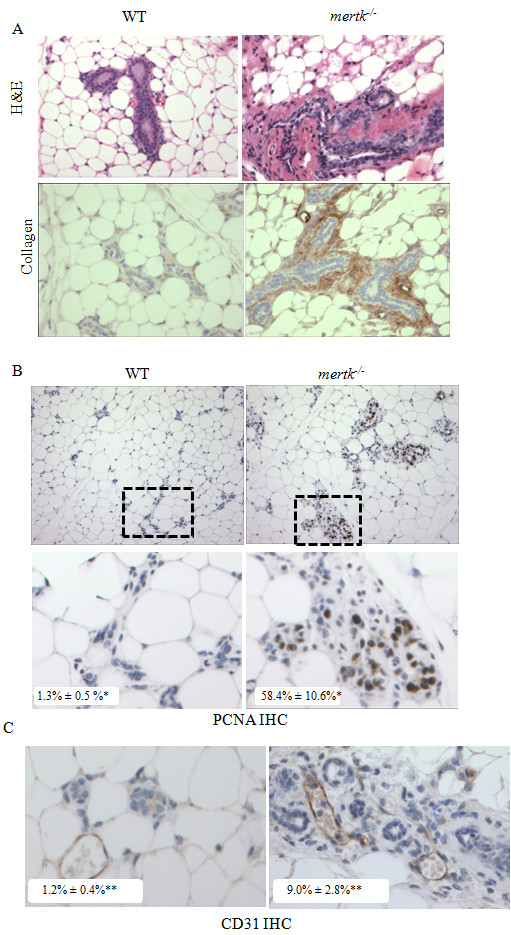
Figure 2.

Impaired apoptotic cell clearance results in pathological changes that are sustained through 60 dpfw. A. Sections of mammary glands harvested at 60 dpfw were stained with H&E and with an antibody against collagen. B. Immunohistochemical detection of PCNA in mammary glands harvested at 60 dpfw. N = 5 per genotype. Values shown in bottom left of each panel represent the average percentage of total nuclei per 400X field (± S.D.) that were PCNA+. *P = 0.045; Student's unpaired T-test, N = 3 samples per group, 5 fields per sample. C. Immunohistochemical detection of the blood vessel marker PECAM/CD31. Values shown in the bottom left of each panel represent the average number of vCD31-positive vessels per 400X field (± S.D.). **P = 0.0001. Student's unpaired T-test, N = 3 samples per group, 5 fields per sample.
