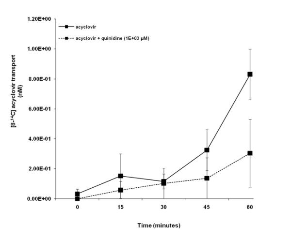
Figure 3.

Acyclovir transport across porcine renal proximal tubular cell (LLC-PK1) monolayers. The transport (basolateral-to-apical) of acyclovir was assessed in LLC-PK1 cells monolayers. Cell monolayers were exposed to [8-14C] acyclovir (5E-02 μM) in the presence or absence of quinidine (1E+03 μM) for 60 minutes. The transport of acyclovir was assessed by measuring the appearance of [8-14C] acyclovir radioactivity in the apical compartment at specific time intervals (0, 15, 30, 45 and 60 minutes) for 60 minutes. Radioactivity was measured as disintegrations per minute (DPM). Acyclovir transport is expressed as the concentration of [8-14C] acyclovir in the apical compartment. Results are presented as the mean (±standard error (SE)) from 3 cell monolayer experiments.
