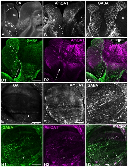
Figure 7. A subset of the GABAergic feedback (PCT) neurons in the mushroom body expresses the AmOA1 receptor.
A: Frontal section of the halves of two calyces labeled with octopamine antiserum (OA). Octopamine immunoreactive processes are in the outer and inner basal ring (o. br, i. br), collar (co) and calyx lip. B: The frontal section adjacent to the section shown in A labeled with anti-AmOA1 antibodies. The AmOA1 positive processes are in the lip, collar and basal ring zones of the calyx in large afferent profiles. Note that the Kenyon cell bodies (K) of the outer basal ring and collar are also labeled with anti-AmOA1. The double arrowheads indicate the outer wedge of the basil ring, which shows relatively intense staining. C: GABA-like immunoreactivity in the feedback neurons have ends in all zones of the calyx. D1-3: Double staining for GABA and AmOA1 in frontal sections of the lateral calyx. Not all of the GABAergic feedback neurons exhibit anti-AmOA1 immunolabeling (arrow). The AmOA1 receptor mostly co-localized with feedback neurons that end in the lip and basal ring zone of the calyx. E: Frontal section through the vertical lobe of the mushroom body labeled with octopamine antiserum. The vertical lobe has octopamine immunoreactive processes only in the γ lobe and in the neuropil of the protocerebrum that surrounds the vertical lobe. F: In a section adjacent to the section shown in E, the vertical lobe of the mushroom body labeled with AmOA1 antiserum. The anti-AmOA1 staining is in: i) extrinsic neurons branching in the γ lobe and ii) processes from feedback neurons that enter the lobe laterally (arrow). The axons of Kenyon cells in the γ lobe labeled with low intensity. G: Frontal section of the vertical lobe labeled with GABA antiserum. The feedback neurons exhibit GABA-like immunoreactivity in profiles that enter in the lobe medio-laterally and branch in the lip, collar and basal ring zone of the vertical lobe. The extrinsic GABA-like immunoreactive processes branch in the γ lobe. H1-3: Double staining with anti-GABA and anti-AmOA1 in the section adjacent to the section shown in G. A subset of GABAergic feedback profiles co-localized with anti-AmOA1. Scale bar: 25 µm.

