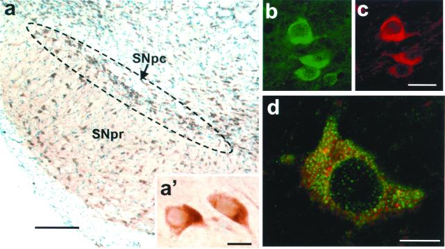
Figure 1.
Bax expression in SNpc dopaminergic neurons of adult mice. (a) Bax is highly expressed in SNpc neurons, as assessed by immunohistochemistry; sections are counterstained with thionin. (a′) High magnification of Bax-immunostained neurons in the SNpc. (b and c) Double immunofluorescence with antibodies to Bax and TH confirms that Bax (in green) is expressed in dopaminergic neurons (in red). (d) Confocal microscopy analysis of Bax-positive dopaminergic neurons (Bax + TH immunostaining) shows a robust punctate immunoreactivity superimposed onto a diffuse cytoplasmic immunostaining. [Scale bars: 200 μm (a), 10 μm (a′ and d), and 30 μm (b and c).]

