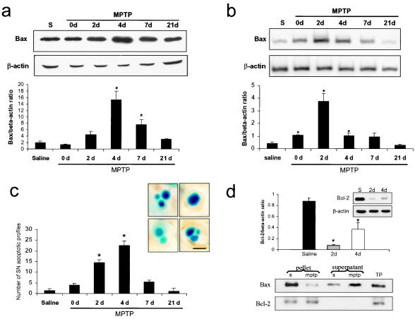
Figure 3.
Bax expression in the ventral midbrain after MPTP intoxication. (a) Bax protein levels in the ventral midbrain (n = 3 mice per group) were assessed by Western blot analysis. (b) Bax mRNA expression in the ventral midbrain was quantified by reverse transcriptase–PCR (n = 3–5 mice per group). (c) Bax protein and mRNA up-regulation coincide with the time course of apoptotic-induced cell death in the SNpc. Morphological criteria to identify apoptotic figures, as illustrated in photomicrographs, included shrinkage of cellular body, chromatin condensation, and the presence of distinct, round, well-defined chromatin clumps, demonstrated by thionin staining. (Scale bar, 5 μm.) (d) Bcl-2 protein expression (Upper) and immunoprecipitation (Lower) after MPTP intoxication. Bcl-2 protein levels are decreased in the ventral midbrain of MPTP-intoxicated mice at days 2 and 4 after the last MPTP injection (n = 3–5 mice per group). At day 4 after the last injection, ventral midbrain proteins (n = 4 mice per group) were subjected to immunoprecipitation with a polyclonal antibody to Bcl-2. The amount of Bax coimmunoprecipitated with Bcl-2 appeared less abundant in the pellets of MPTP-intoxicated mice than in those of saline-injected animals. This was associated with increased Bax immunoreactivity in the supernatant. S, saline; TP, total proteins. *, P < 0.05, compared with saline-injected animals; Newman-Keuls post hoc analysis. Error bars indicate SEM.

