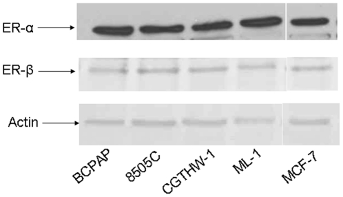
Figure 1. Thyroid cells express estrogen receptor.
Whole cell protein (20 µg) was resolved by SDS-PAGE followed by Western blot analysis for ER-α (dilution 1∶500), ER-β (dilution 1∶1000) and actin (dilution 1∶5000). All the cell lines used in this study (BCPAP, 8505C, CGTHW-1 and ML-1) express both ER-α and ER-β at comparable levels.

