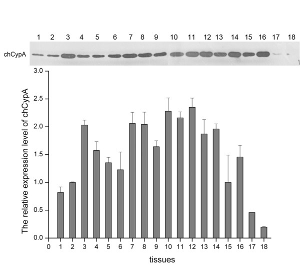
Figure 5.

The distribution of chCypA in different tissues. Eighteen tissues were extracted from 21-day-old SPF chickens and relative protein level of chCypA was determined by Western blot and relative densitometry analysis carried out with Photoshop program. 2 μg tissues extracts were loaded into SDS-PAGE. 1, heart 2, liver, 3, spleen 4, lung 5, kidney 6, pancreas 7, brusa of Fabricius 8, esophagus 9, duodenum 10, thymus 11, cerebrum 12, cerebella 13, glandular stomach 14, gizzard 15, muscle 16, trachea 17, ovary 18, blood. Tissues of three chickens had been extracted, and the representative data is shown.
