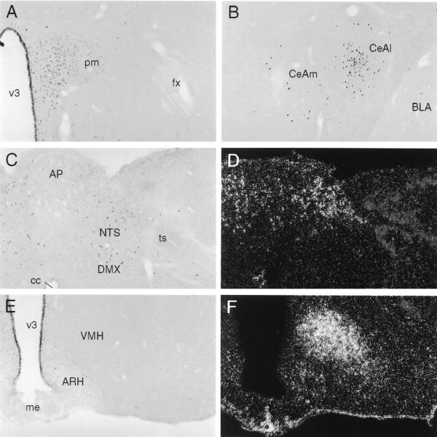
Figure 3.
Cellular activation patterns in response to central Ucn II microinjection. (A-C and E) Brightfield photomicrographs of immunoperoxidase preparations showing induced Fos expression in rats killed 2 h after i.c.v. injection of 1 μg of synthetic mouse Ucn II. Darkfield photomicrographs showing hybridization histochemical localization of CRF-R2 mRNA in regions corresponding to those illustrated in C and E are provided in D and F, respectively. Central Ucn injection provoked Fos induction primarily in a set of interconnected structures involved in central autonomic and neuroendocrine control, including the parvocellular division of the paraventricular nucleus (A), the central nucleus of the amygdala (B), and the nucleus of the solitary tract (NTS, C). Among these, only the NTS is a site of CRF-R2 expression (D). Other principal sites of CRF-R2 expression, including the ventromedial nucleus of the hypothalamus (F), failed to show Ucn II-induced Fos expression over the range of peptide doses examined (1–10 μg). (Magnification for all photomicrographs: ×75.)

