
Figure 2.
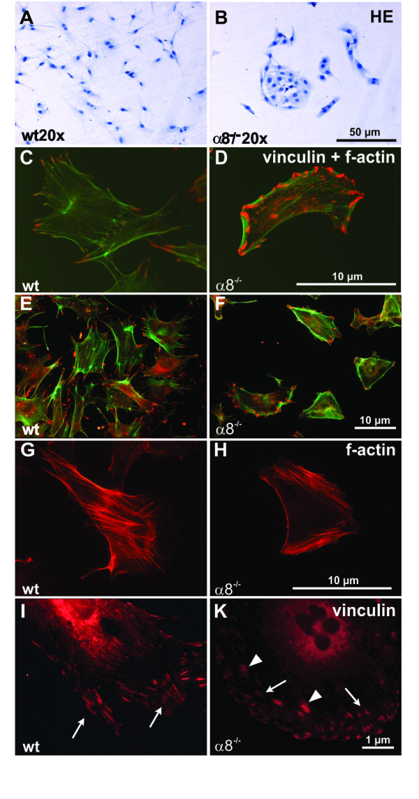
Comparison of wild type (wt) and α8 integrin-deficient (α8-/-) mesangial cell morphology after hematoxylin stain (A+B), immunofluorescent double staining for f-actin in green and vinculin in red (high magnification C+D, low magnification E+F), immunofluorescent staining for f-actin alone (G+H) or immunofluorescent staining for vinculin alone (I+K). White arrows indicate focal contacts of the cells and white arrowheads indicate bundles of focal contacts in α8-/- mesangial cells.