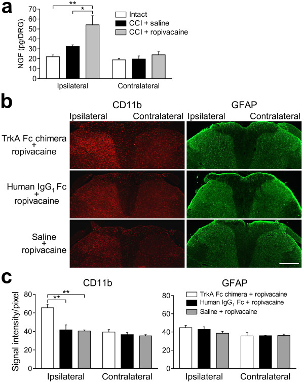
Figure 3.
Effects of the TrkA-Fc chimera on the ropivacaine-induced suppression of activated glial cells in CCI rats. (a) The NGF contents on the contralateral and ipsilateral sides were measured in control intact rats (white columns), CCI rats treated with epidural saline (black columns) and CCI rats treated with epidural ropivacaine (grey columns). *p < 0.05 and **p < 0.01 by Tukey-Kramer's multiple comparison test (n = 8-9). (b) Representative images of immunohistochemical staining for CD11b (a microglial activation marker) and GFAP (an astrocyte activation marker) in the L4 spinal dorsal horn. L4 spinal cords were obtained from CCI rats treated with the TrkA-Fc chimera and ropivacaine, CCI rats treated with the control IgG1-Fc protein and ropivacaine, and CCI rats treated with saline and ropivacaine at day 10 after CCI. Scale bar, 200 μm. (c) Densitometric quantification of the CD11b and GFAP immunoreactivities. **p < 0.01 by Tukey-Kramer's multiple comparison test (n = 4).

