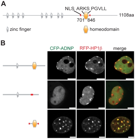
Figure 4. The homeodomain of ADNP is necessary and sufficient for localization to pericentromeric heterochromatin.
(A) Schematic representation of ADNP protein structure (Uniprot ID Q9H2P0). Positions of the nine zinc-fingers (grey), the homeodomain (orange), the nuclear localization signal (NLS) and the ARKS and PGVLL motifs are indicated. (B) NIH3T3 cells were transfected with CFP-ADNP, CFP-ADNP(D741-846) (homeodomain deletion), CFP-ADNP(701-846) (NLS and the homeodomain) and RFP-HP1β. Only cells of low to medium expression levels showed distribution of wt CFP-ADNP similar to the endogenous protein. Therefore, the images show CFP and RFP fluorescence signals of selected unfixed, living cells of low expression levels of the different CFP-fusion proteins using a confocal microscope. Bars, 5 µm.

