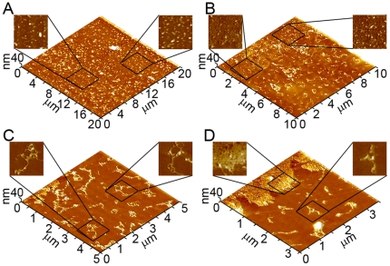
Figure 6. Representative in situ AFM images of Italian Aβ (1–40) aggregate formation on supported TBLE lipid bilayers.
(A) Upon addition of a 20 µM solution of Italian Aβ (1–40) to the TBLE bilayer, oligomeric aggregates appeared within 2–4 hours. (B) After ∼10–12 hours of exposure to Italian Aβ (1–40), the bilayer developed large patches of increased bilayer roughness that often contained oligomeric aggregates. (B–D) Elongated fibrillar aggregates were observed within 8–10 hours of addition of Italian Aβ (1–40) to the lipid bilayer. These fibrillar aggregates displayed a variety of morphologies, predominantly displaying large curvature and branching.

