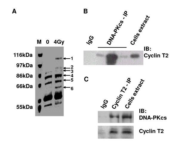
Figure 7.
The interaction of DNA-PKcs with cyclin T2. A: Coomassie bright blue dye staining of SDS-PAGE of the co-immunoprecipitation products of DNA-PKcs antibody. B: Immunoblotting analysis of cyclin T2 was performed on the CoIP products of DNA-PKcs antibody and total extracts of cells. Three DNA-PKcs lanes represent three independent repeat IP experiments. C: Immunoblotting analysis of DNA-PKcs and cyclin T2 was performed on the CoIP product of cyclin T2 antibody and the total extracts of cells. The immunoprecipitation product of Ig G was taken as the blank control.

