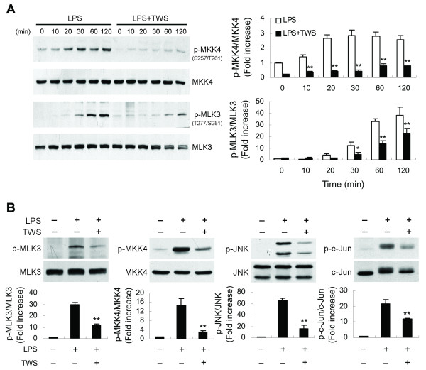
Figure 7.
Decreased GSK-3β activity blocks MLK3 signaling. BV-2 cells (A) or primary microglia (B) were pretreated with vehicle or TWS119 (2.5 μM) for 30 min and then stimulated with LPS (100 ng/ml) for the indicated times (A) or 30 min (B). Western analysis was used to determine total and phosphorylated c-Jun, JNK, MKK4 and MLK3 proteins in whole cell extracts. Data are presented as mean ± SEM for three independent experiments. **p < 0.01 compared with respective cultures treated with LPS alone.

