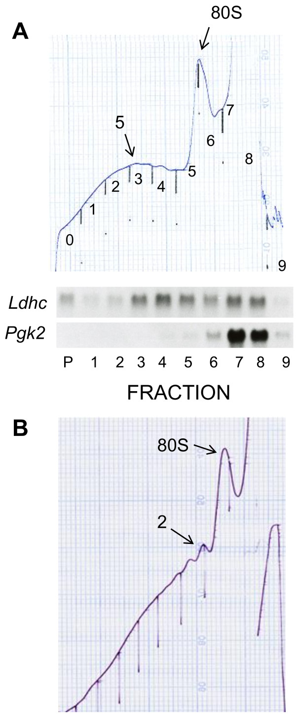
Figure 2.
Analysis of mRNA translation in purified pachytene spermatocytes purified by sedimentation on bovine serum albumin gradients. Dissociated testicular cells from 16 dpp (A) and adult mice (B) were purified by sedimentation at 1XG on bovine serum albumin gradients, pachytene spermatocytes were collected, cultured for 1 hr at 32°C in RPMI 1040 medium in 5% CO2 in air, cytoplasmic extracts were prepared and sedimented on sucrose gradients. The preparation in panel A contained 4.28 × 10⁶ pachytene spermatocytes. The gradients in panels A and B were sedimented at 35,000 rpm in the SW60Ti rotor for 100 and 80 min, respectively. The absorbance tracings of both gradients at 254 nm are shown. In addition, in Panel A the RNAs were extracted from each fraction, and the distribution of the Ldhc and Pgk2 mRNAs was analyzed by sequential hybridization of a single Northern blot. The fractions were collected from the bottom, and the fraction labeled P contains RNA extracted from the pellet on the bottom of the ultracentrifuge tube. The full scale absorbance of the UV analyzer was 0.32 using a flow cell with a 2 mm path length. The positions of the fractions in the absorbance tracing in panel B are numbered. Fraction 0 in the absorbance tracing is the void and does not contain RNA.

