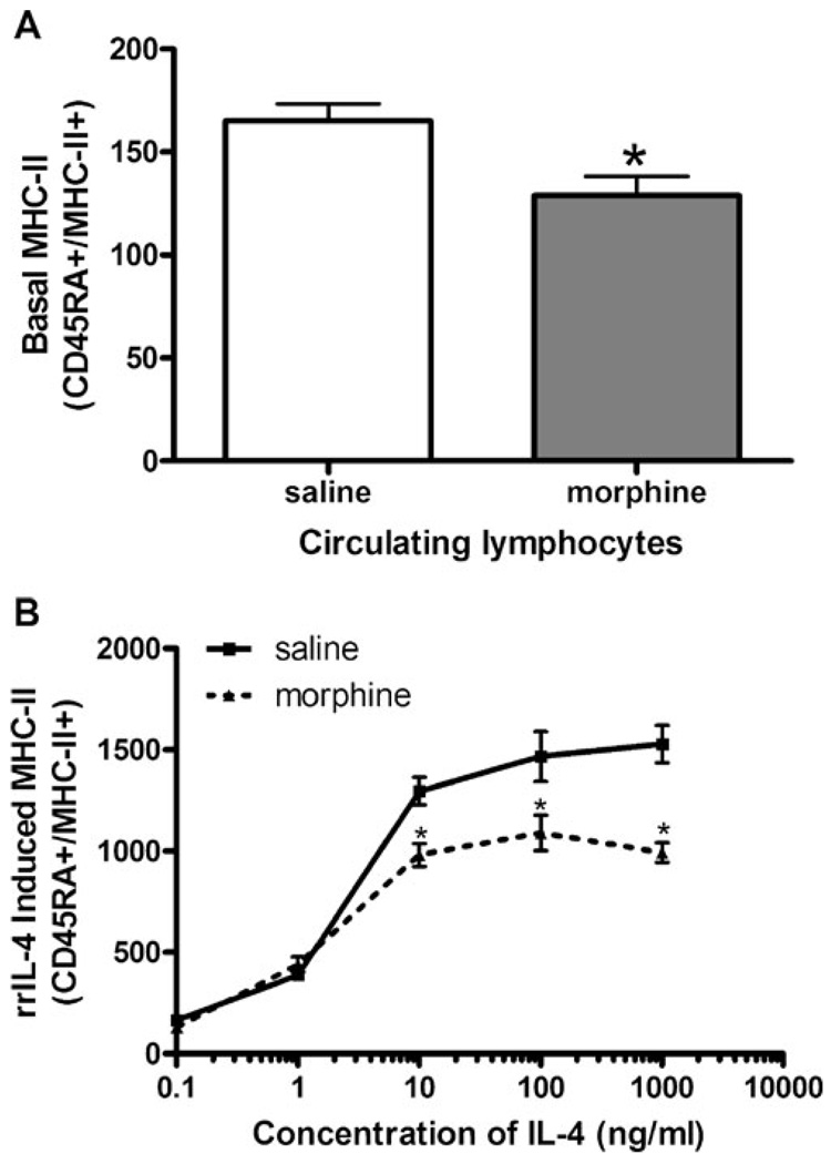
Fig. 1.
Basal MHC-II expression after treatment with a single dose of morphine in circulating B lymphocytes. a Groups of Sprague–Dawley rats (n=6) were administered either saline or morphine (10 mg/kg, s. c.). Two hours after morphine injection, the rats were rapidly decapitated and peripheral blood mononuclear cells were isolated from whole blood. Leukocytes were isolated and incubated with fluorochrome-conjugated antibodies as follows: anti-rat CD45RA-PE (lymphocytes) and anti-rat MHC-II RT.1B β FITC (MHC-II). Stained cells were fixed using 1% paraformaldehyde prior to analysis using a FACSort flow cytometer. Data are representative of three separate experiments and are expressed as MFI ± SEM. *p<0.05 compared to all other treatment groups (Student’s t test). b rrIL-4 to induce MHC-II expression in B lymphocytes after a single injection of morphine. Sprague–Dawley rats (n=6 per group) were administered either saline or morphine (10 mg/kg, s.c.). Two hours after the subcutaneous injection, peripheral blood mononuclear cells were isolated and cultured at 37°C and 8% CO2 with rrIL-4 (0.1–1,000 ng/ml). Cells were collected and washed prior to being incubated with fluorochrome-conjugated antibodies, as described above. Fixed cells were analyzed using a FACSort flow cytometer. Data are expressed as MFI ± SEM. Data repeated in two subsequent experiments. *p<0.05 compared to control (ANOVA, Newman–Keuls)

